
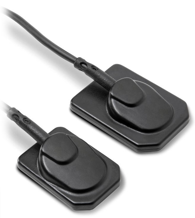
I-View Gold
Trident SRL · source
I-VIEW GOLD - INTRAORAL CMOS SENSOR
I-View Gold covers a wide range of exposure settings, capturing a consistent and diagnosable image under any conditions.
Combine I-View Gold with your traditional X-ray generator and obtain incredibly detailed images.
- TWAIN interface
- DICOM files format
- Bridge included (this function links the sensor to the management software)
HIGH-DEFINITION DIGITAL IMAGES
I-View Gold captures images of the most critical areas of the oral cavity both horizontally and vertically, with a resolution of 27 lp/mm visible and a pixel size of 18 µm.
LARGER ACTIVE SURFACE
Active Pixels
- Size 1: 1110×1666 pixels
- Size 2: 1442×1998 pixels
MULTIUSER CONNECTION
The software can be installed on several computers. Up to ten users on the network simultaneously.
THINNER BODY
I-View Gold is much more comfortable for your patients, it is only 4.8 mm thickness.
DIRECT USB – COMPUTER CONNECTION
I-View Gold intraoral sensor is directly connected to your PC with a 2,7-metre USB cable (connector compatible with a standard 2.0 USB port). The sensor is used as a temporary device and is not connected to the mains power supply; power is provided through the computer.
WATER RESISTANT
IP Code: 68. The sensor was designed to withstand dust, dirt and sand, and is resistant to submersion underwater.
HIGH-DEFINITION IMAGES WITH NO FILTERS
Thanks to a highly evolved calibration tool, images obtained with I-View Gold do not need post-processing: during the acquisition phase the calibration files remove any possible imperfection by reducing noise and increasing details.
Two sizes
Rounded corners
- Flat smooth surface
- Silicon protective cover
- Active Area:
- Size 1: 20×30 mm
- Size 2: 26×36 mm
I-VIEW GOLD: ULTIMATE DURABILITY & EFFORTLESS INTEGRATION
Reliable & Secure
Stable USB 2.0 Connection. Plug Directly into Your Computer for instant use.
Built to endure
IP68-rated – fully waterproof and submersible in liquids. Resistant to dust and extreme cold for maximum reliability. Designed to perform in any environment.
Deep-View software for intraoral images
Deep-View is a modern imaging software designed by Trident to efficiently acquire, organize, store and share digital images. The suite is a complete and integrated software for intuitive navigation with several advanced features to obtain and manage thousands of high-definition images for more accurate diagnosis.
EFFICIENT
Save time, optimize files' management, and speed up the workflow.
EASY TO USE
Run the amazing features of Deep-View in a few simple steps.
MULTIPLATFORM
The interface innovative design makes Deep-View easy to use in multiple devices.
SECURE
Advanced cryptography techniques ensure data protection.
SENSOR HOLDER
Ensures stable positioning of the intraoral sensor, making placement in the patient's mouth easier and more precise. The result: sharp, high-quality images in less time.
- Ergonomically designed for greatest patient comfort.
- Securely holds sensor in place while giving you the precision and control you need to obtain the right shot.
- Available in two sizes, size 1 and size 2, in bright and vivid colors for easy identification.
- One ring, no changing between different rings for specific images.
- Just by rotating the sensor holder, it is possible to use it in the upper and lower jaw.
KIT CONTENT
1 X POSITIONING RING
The ring offers a perfect fixation and alignment of the holder. To protect the sensor cable, the positioning ring is equipped with cable guides.
2 X PERIAPICAL SENSOR HOLDER
- 1 model for upper left and lower right (UL/LR)
- 1 model for upper right and lower left (UR/LL)
2 X ENDO SENSOR HOLDER
- 1 model for upper left and lower right (UL/LR)
- 1 model for upper right and lower left (UR/LL)
2 X POSTERIOR SENSOR HOLDER
- 1 model for upper left and lower right (UL/LR)
- 1 model for upper right and lower left (UR/LL)
1 X BITEWING HOLDER
The bitewing exposures using the Bitewing sensor holder, result in images in which the premolar regions are clearly visible. Thanks to the good position of the sensor the image covers a considerably larger area. The interdental spaces are sharply focused.
Informations produit
| Fabricant | Trident SRL |
|---|---|
| Ip Rating | IP68 |
| Multiuser | Up to ten users on network simultaneously |
| Connexion | TWAIN |
| Pixel Size | 18 µm |
| Résolution | 27 lp/mm visible |
| File Format | DICOM |
| Sensor Type | Intraoral CMOS Sensor |
| Cable Length | 2.7 metres |
| Alimentation | Via USB |
| Body Thickness | 4.8 mm |
| Water Resistance | Waterproof and submersible |
| Included Software | Deep-View |
| Active Area Size 1 | 20×30 mm |
| Active Area Size 2 | 26×36 mm |
| Active Pixels Size 1 | 1110×1666 pixels |
| Active Pixels Size 2 | 1442×1998 pixels |