
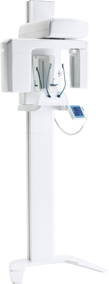
X-VIEW 2D PAN
Trident SRL · source
The latest technology for perfect images with high diagnostic value.
Thanks to the introduction of the most recent technology and the newest features in digital radiology, X-VIEW 2D PAN requires a minimum exposure time to capture clear and contrasted images.
Panoramic images acquired with X-VIEW 2D PAN have a precise focus throughout the entire arch and facilitate the general evaluation of the quantity and quality of bone, dentition and TMJ.
FEATURES
HIGH FREQUENCY DC X-RAY GENERATOR
For images with low noise and reduced radiation dose to the patient. Exposure time: 2.4-15.5 s
FOCAL SPOT: 0.5 mm
CMOS SENSOR FOR PAN
- Area: 150×6.9 mm
- Pixel size: 100 µm
- Number of pixels: 1500×68
- Higher Sensitivity
- Lower dark noise
- High Pixel well depth
- Lower Power consumption
- Free from smear
7-INCH COLOR TOUCHSCREEN
INTUITIVE GRAPHICAL USER INTERFACE
All instructions available in a number of languages allow users to easily access the different options
DR CMOS SENSOR FOR CEPH
- Better contrast
- More details and filtering
- No background disturbance
- Calibration method: easy intuitive and manageable from remote
MADE IN ITALY
Elegant and compact Italian design with a space-saving structure
QUICK AND EASY PATIENT POSITIONING
The ergonomic design ensures that the patient is not enforced to use awkward postures and can easily achieve the correct positioning.
The specially designed tools assist patients, during the acquisition process, to maintain their position avoiding errors in the procedures.
X-VIEW 2D adapts to all sizes and types of patients, the linear and open design facilitates access for wheelchair users.
7-INCH COLOUR TOUCHSCREEN
The LCD display enables users to directly interact with the unit.
Menus are very easy to navigate.
Universally recognized icons indicate available functions, parameters, and options.
All instructions are multilingual in a friendly language that guides the user fluently through the different options.
Available in three models to offer complete and affordable imaging solutions:
X-VIEW 2D PAN BASIC
This is the simplest model of the X-VIEW family, developed to ease doctors switching from an intraoral to an extra-oral digital imaging system.
- Easy installation
- Low maintenance
- Low initial investment cost
- Easy to use
CMOS SENSOR FOR PANORAMIC IMAGES:
- 30×15 cm adult panoramic in 15.5 s
- 30×13 cm child panoramic in 14.8 s
X-VIEW 2D PAN UP
This model grows along with the workflow. Initially the doctor acquires an X-VIEW 2D PAN UP that in a second stage becomes a 2D PAN CEPH.
X-VIEW 2D PAN UP, born as a 2D PAN BASIC model with the specific mechanical and user interface dispositions to articulate the cephalometric option in a second stage.
The upgrade process consists of adding the cephalometric arm and a dedicated DR single shot sensor for cephalograms.
X-VIEW 2D PAN CEPH
The 2-in-1 model makes the advantages of 2D and cephalometric imaging within reach of the general dentists, periodontists, orthodontists and other specialists working from medium offices to hospitals and clinics.
TWO DETECTORS
In addition to the CMOS detector for all 2D images, X-VIEW 2D PAN CEPH is equipped with a dedicated DR flat panel detector for Cephalograms acquisition. The DR (Digital Radiography) version is the highest quality technology available to obtain sharp Ceph images.
SAME SOFTWARE AND DATABASE FOR BOTH 2D AND CEPH PROGRAMS
FULL CONNECTIVITY TO EASILY EXPORT, SHARE AND STORE IMAGES
- Variable field collimator
- 30×15 cm adult panoramic in 15.5 s
- 30×13 cm child panoramic in 14.8 s
- 24×30 cm AP/LL cephalograms
- Carpus images
FUNCTIONS
2D PAN FUNCTION
The standard panoramic program provides a precise definition of the dental anatomy in just 15 seconds allowing:
- Overall evaluation of dentition
- Examine for intraosseous pathology, such as cysts, tumors, or infections
- Gross evaluation of temporomandibular joints
- Evaluation of position of impacted teeth
- Evaluation of eruption of permanent dentition
- Dentomaxillofacial trauma
- Development of disturbances on maxillofacial skeleton
CEPH FUNCTION
24×30 cm AP, LL cephalograms
Carpus images
- Describes the patient's dento-facial morphology
- Quantitatively describes the morphological deviations
- Obtains orthodontics diagnosis and treatment planning
- Supports the planning of maxillofacial surgery
- Elaborates Airway analysis
- Analyzes the skeletal and dental abnormalities
- Evaluates treatment results
- Predicts growth related changes
GO TO THE NEXT LEVEL WITH FOCUS
Enhance your professional practice with cutting-edge technology in digital 2D image acquisition. The new X-VIEW 2D PAN FOCUS, equipped with advanced Image Reconstruction Technology, surpasses traditional methods for capturing panoramic images.
FOCUS technology generates high-definition 2D images derived from CBCT technology, applying tomographic techniques to the OPG field. These newly acquired images are exceptionally sharp and clear, eliminating the need for post-processing.
During acquisition, the autofocus function automatically identifies the optimal focus zones, ensuring the best possible sharpness and resolution—delivered quickly and with lower radiation exposure.
The X-VIEW 2D PAN FOCUS meets the highest expectations of healthcare professionals. Its dynamic, user-friendly design and flexible technology consistently deliver excellent results, even in the most complex cases.
FUNCTIONAL OVERVIEW
The FOCUS technology is purely based on mathematical operations and the proper calibration of the reconstruction parameters during the X-ray system development phase. The determination of both a proper system and the reconstruction parameters, ensures that a volume of planes spans all the parts of the patient's relevant anatomy with planes dense enough to ensure that any part of the relevant anatomy is captured with adequate sharpness at least in one of the planes in the reconstructed stack.
- PROJECTION FRAME - Individual images acquisition from the detector with offset, gain and defect pixel correction.
- TOMOGRAPHIC PLANE RECONSTRUCTION - A set of tomographic planes is created by combining the individual projection frames through a so-called "shift-and-add" method. This process is performed in parallel for several focal planes, using different pixel shift values.
- ANATOMY CONDITIONING - Clinically irrelevant features in select tomographic planes can be suppressed, to reduce their visibility in the final reconstructed image.
- 2D IMAGE SYNTHESIS - From the full set of (conditioned) tomographic planes, a single 2D image is synthesized that combines the sharpest information from the total set of tomographic planes throughout different regions of the image. Vertically, regions from planes at different depth positions can be combined to follow complex patient anatomy (e.g. overbite).
- GEOMETRY CORRECTION - The 2D reconstructed image can be scaled in the scan direction, to adjust for magnification and scaling differences due to system geometry and scan motion profiles.
Informations produit
| Fabricant | Trident SRL |
|---|---|
| Affichage | 7-inch color touchscreen |
| Focal Spot | 0.5 mm |
| Résolution | 100 µm |
| Place Of Origin | Fabriqué en Italie |